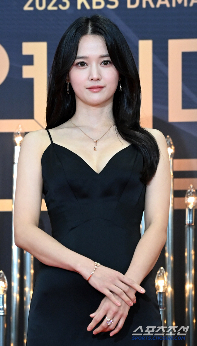
80% of premature infants born before 28 weeks have retinopathy, and if left unattended, there is a possibility of causing blindness

Feb 05, 2025

80% of premature infants born before 28 weeks have retinopathy, and if left unattended, there is a possibility of causing blindness
data photo source=Pixabay


Since the formation of blood vessels in the retina progresses slowly from 4 months to about 40 weeks of birth, the retina of premature babies usually grows outside the mother's body. At this time, if blood vessels do not grow to the end due to changes in the environment or new blood vessels grow together, 'immature retinopathy' may occur.

With recent advances in medicine, the number of ultra-premature infants under 25 weeks of gestation has increased, and the occurrence of 'immature retinopathy' is also increasing. Professor Kim Jin-ha of Soonchunhyang University Bucheon Hospital summarized 'immature retinopathy'.

Professor Kim Jin-ha "Domestic studies have reported that retinopathy in premature infants occurs in about 30% of premature infants under 37 weeks of gestational age. Early detection and treatment are the most important in preventing blindness, so screening tests for retinopathy of premature infants should be performed for all premature infants."


◇About 80% of pre-28 weeks' birth 'immature retinopathy' found

Prenatal retinopathy occurs because the environment inside the mother's uterus and outside the mother is different. When born, the physiological environment supporting blood vessel formation is cut off, and oxygen partial pressure difference occurs, causing blood vessels to not grow properly, blood vessel obstruction, or vascular proliferation, resulting in retinopathy of premature infants.

The major risk factors for retinopathy in premature infants are gestational age and birth weight. The lower the gestational age and birth weight, the higher the incidence of retinopathy in premature infants, the more severe the degree, and the faster the progression. According to the CRYO-ROP study, infants born after 32 weeks have little retinopathy of premature infants, but about 60% of infants born at 28-31 weeks and 80% of infants born before 28 weeks have retinopathy of premature infants. In addition, there is a significant difference in prevalence, with a weight of less than 1500g being about 30%, a weight of less than 1500g being about 2.5%, and a weight of more than 2500g being 0.03%.


◇ Observe the retinal vascular formation region and vascular changes with fundus examination

To evaluate whether prematurity retinopathy is present, a fundus examination is performed. Since the fundus examination should be carried out with sufficiently large pupils, check with an ophthalmoscope after the eyeball of Sandongje. The patient's eyes are fitted with a lidar, and the scleral press is used to observe changes in the surrounding retina, including the optic papilla and the posterior retinal blood vessels. With the help of the intensive care unit, the fundus test closely monitors changes in the child's whole body condition and proceeds so as not to be burdened with premature babies.

Professor Jinha Kim observes and evaluates retinal vascular formation regions and vascular changes through this. The wider the area where blood vessels grow, the better the prognosis, and the narrower the area, the higher the need for treatment. In addition, if there is a boundary between the vascular formation site and the vascular intangible site, or if intravascular shortcuts, extraintestinal fibroblasts, vitreous proliferation, and retinal detachment are observed, they must be treated."


◇ When left unattended, complications such as scarring or retinal detachment causing blindness are accompanied

Prenatal retinopathy can be accompanied by serious complications such as spontaneous regression, scarring, or retinal detachment leading to blindness. The purpose of treatment is to prevent the development of new blood vessels, scarring, and complication of retinal detachment in areas where blood vessels have not grown.

In the past, cryotherapy was standard treatment, but recently laser therapy has become a representative treatment. After local or general anesthesia, the laser is irradiated to the vascular intangibles without gaps. After that, loss such as optic papillary change, vascular change, and bleeding occurs, stabilizing the retina and strengthening the adhesion of the vascular-free retina, it is considered a successful treatment.

Professor Kim Jin-ha "However, despite successful laser treatment, some may progress to retinal detachment. In this case, surgery such as sclerodolectomy or vitreous resection should be performed depending on the degree of retinal detachment. In addition, there may be complications such as refractive abnormalities, cataracts, decreased vision, and strabismus, as well as anatomical changes such as macular drag and macular fold."

Recently, attempts have been made to treat retinopathy in premature infants 'injections of anti-vascular endothelial growth factors in the free body cavity (injections in the free body cavity).' Studies have also been published that intra-glass lumen injections are relatively safe and have less incidence of myopia and other refraction abnormalities than laser treatment groups. However, studies have also reported that such injection therapy is temporary and can worsen the risk of recurrence, systemic side effects, and retinal detachment, so it should be applied carefully.

Professor Kim Jin-ha said "The incidence of prematurity retinopathy is decreasing due to the improvement of premature infant care and advances in treatment research over the past decades. Since the major risk factors for retinopathy in premature infants are gestational age and weight, it is important to get help to have a healthy birth in consultation with your obstetrician if there is a risk of premature birth or a risk of low birth during pregnancy. After birth, early detection and treatment through appropriate screening tests are the key."

80% of premature infants born before 28 weeks have retinopathy, and if left unattended, there is a possibility of causing blindness
김진하 교수


This article was translated by Naver AI translator.